Dr. Maren Strenziok, a post-doctoral fellow at George Mason University's Arch Lab presented the research that she and numerous other faculty and students are currently involved in. The backbone of their research consists of conducting training and longitudinal studies in healthy aging. These studies are vital as they attempt to make sense of why the brain undergoes decline with age. Decline begins at the age of 30-40 years and is steady in terms of brain tissue integrity. On average, cognitive function and fluid abilities and intelligence decline, but this does not occur in everyone. About 30-50% of people show no decline in cognitive function. Different factors including diet, exercise, education, and genetic and social factors are studied for any correlation and to help explain this phenomenon (2).
 |
This image is of a diffusion tensor imaging scan. DTI scans of a healthy brain add color to its white matter. http://www.dana.org/news/brainwork/detail.aspx?id=5322 |
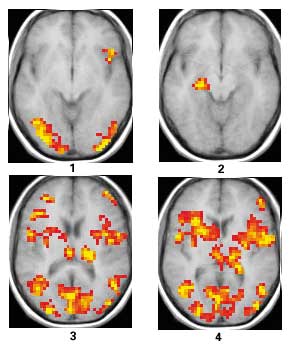
Brain aging has been studied using magnetic resonance imaging (MRI). Dr. Strenziok introduced the techniques used and focused mainly on two types of MRI techniques: diffusion tensor imaging (DTI) and functional magnetic resonance imaging (fMRI). DTI is an MRI technique that allows for the measurement of fractional anisotropy, the random diffusion of water molecules in the brain. Higher fractional anisotropy (FA) reflects better white matter fiber integrity while a decline in FA is indicative of decreasing white matter health. Functional MRI is a technique that helps in mapping the specific regions of the brain engaged in certain tasks, processes or emotions. It works by detecting changes in blood flow related to neural activity of brain cells. It was used to measure the functional connectivity of the brain when in default mode network (DMN).
 |
| This image depicts four fMRI brain scans obtained during a visual memory task. http://berkeley.edu/news/media/releases/2000/11/20_mri.html |
In the study conducted at the Arch Lab, a video game known as 'Rise of Nations' was used. This video game was previously shown to have benefited older people and an increase in white matter integrity and improved intellectual ability have been noted. Training was conducted in individuals over the age of 60 and the study lasted 8 weeks. Participants trained 6 times a week; 3 times were supervised in the lab and 3 times were at home. Each session lasted for 1 hour. There were 10 trained subjects and 8 control subjects. Experimenters engaged in motivational speech with the participants. A fixed order of games was used and an emphasis was placed on using a variety of strategies. Pre and post-training performance differences were measured across participants (2).
 |
| Rise of Nations: Rise of Legends Preview http://pcmedia.gamespy.com/pc/image/article/685/685658/rise-of-nations-rise-of-legends-20060203034016276-000.jpg |
The results of the experiment were not as expected. Corpus callosum FA was lower post-training in trained individuals. This was a surprise as white matter integrity is expected to increase and a greater FA should mean better performance. This is not always the case however as was found in a study in which Williams Syndrome patients were tested. Although these participants were found to have higher FA post-training, they did worse on tests. It can be concluded therefore, that FA can decrease with expertise, as well as with age. In functional connectivity tests, superior parietal connectivity was higher post-training in trained individuals. It was noted that participants exhibited more reliance on intrahemispheric integration and less reliance on interhemispheric integration. A correlation was also made between lower FA and higher functional connectivity in the same participants. In the future, the team at Arch lab hope to create an index to measure improvement over time in game performance (2). They also hope to further analyze cortical thickness and how it degenerates with age.
Overall, I found Dr. Strenziok's lecture to be informative and thought-provoking and her presentation style was also effective. The research that is being conducted at the Arch lab is vital and can help in better understanding abnormalities of the brain associated with different psychiatric disorders such as obsessive compulsive disorder and depression. By providing insight into the abnormalities and faulty neural connections associated with such disorders, techniques such as fMRI and DTI can provide clues to recovery. By employing these techniques in people at risk but not yet suffering from a developmental disorder, it might possibly enable treatment before the onset of symptoms (3). Patients with traumatic brain injuries (TBI) have symptoms that manifest themselves in different ways. Would it be possible to use fMRI scans to predict how patients with TBI will respond to different rehabilitation strategies?
References:
1. http://www.nursingassistantcentral.com/blog/2008/100-fascinating-facts-you-never-knew-about-the-human-brain/
2. Strenziok, Dr. Maren. "Brain Connectivity: Neural Substrates of Cognitive Function in Healthy Aging", Arch Lab/Cognitive Genetics Research Group – Psychology. 20 September 2012. Lecture.
3. http://www.dana.org/news/brainwork/detail.aspx?id=5322&p=1
4. http://www.dana.org/news/publications/detail.aspx?id=14442

